Cross Section Of A Bone Diagram / How to draw the diagram of cross section of a leaf class x.. We don't draw the rest of the object, just the shape made when you cut through. Cross section of bone diagram. Create your own flashcards or choose from millions created by other students. Cross section of the long bone. Each system contains for a bone tissue engineering scaffold to be successful, it must be highly porous, osteoconductive, biodegradable, biocompatible, mechanically.
Explaned distal and proximal epiphysis. This bone is located directly beneath the skin on the anterior aspect of the leg (top of the image). Histology sauropod vertebra picture of the week these pictures of this page are about:long bone cross section. Jump to navigation jump to search. Bone is hard and many of its functions depend on that characteristic hardness.
Plos One Spectroscopic Studies On Organic Matter From Triassic Reptile Bones Upper Silesia Poland from journals.plos.org A cross section of a human long bone. Bone marrow is the soft, highly vascular and flexible connective tissue within bone cavities which serve as the primary site of new blood cell production or bone marrow is the primary source of pluripotent stem cells that give rise to all hemopoietic cells (blood cells) including lymphocytes. Some descriptions for confusing partsomit number 13 in the picture. The surface features of bones vary considerably, depending on the function and location in the body. Histology sauropod vertebra picture of the week these pictures of this page are about:long bone cross section. Explaned distal and proximal epiphysis. For example, to read this diagram literally, since the cartilage can be seen inside the cutaway section of. Two types of bone tissues in cross section of a long bone :
Cheek bone (zygoma) upper jaw.
From wikimedia commons, the free media repository. 12 photos of the cross section of human bone diagram. We can see there are two layers of compact bone here. Cross section of bone diagram. Find the perfect bone diagram stock illustrations from getty images. Related posts of cross section of human bone diagram bones of the left ankle with diagram. Grossly, bone tissue is organized into a variety of shapes and configurations adapted to the function of each bone internal structure of a human long bone, with a magnified cross section of the interior. Human respiratory system anatomical line style artistic vector illustration, medical education cross section diagram. Diagram with articular cartilage, marrow, spongy bone, medullary cavity, endosteum, diaphysis, and. Spongy bone diagram schematic diagram. The cross section of a rectangular pyramid is a rectangle. Explaned distal and proximal epiphysis. How to draw the diagram of cross section of a leaf class x.
Human respiratory system anatomical line style artistic vector illustration, medical education cross section diagram. The diagram of a long bone could become your choice when making about bone. How to draw the diagram of cross section of a leaf class x. Bone marrow is the soft, highly vascular and flexible connective tissue within bone cavities which serve as the primary site of new blood cell production or bone marrow is the primary source of pluripotent stem cells that give rise to all hemopoietic cells (blood cells) including lymphocytes. Some descriptions for confusing partsomit number 13 in the picture.
Bone Cross Section Photos And Premium High Res Pictures Getty Images from media.gettyimages.com I am not an expert on this subject, so i was wondering if i don't like way you've shown the cartilage. As shown in figure 2. Whereas a long bone has only one layer of compact bone (see fig 1). Jump to navigation jump to search. The periosteum contains many strong collagen fibers that are used to firmly anchor. Diagram of blood and nerve supply to bone. Cross section of the long bone. This bone is located directly beneath the skin on the anterior aspect of the leg (top of the image).
Create your own flashcards or choose from millions created by other students.
The diagram of a long bone could become your choice when making about bone. Explaned distal and proximal epiphysis. The periosteum contains many strong collagen fibers that are used to firmly anchor. The centroidal distance, c, is the distance from the centroid of a cross section to the extreme fiber. The surface features of bones vary considerably, depending on the function and location in the body. Cross sections are usually parallel to the base like above, but can be in any direction. How to draw the diagram of cross section of a leaf class x. Two types of bone tissues in cross section of a long bone : Grossly, bone tissue is organized into a variety of shapes and configurations adapted to the function of each bone internal structure of a human long bone, with a magnified cross section of the interior. Blood vessels and nerves enter the bone through the. □ on examining a cross section of any bone, it is composed of two kinds of bony tissue: Cross section of the long bone. This bone is located directly beneath the skin on the anterior aspect of the leg (top of the image).
Create your own flashcards or choose from millions created by other students. In the last decade, considerable technological improvements have been made to repair damaged bones and tissue, such as bone cross sections with implants for microscopic examinations. Diagram of a male upper leg. Looking at a bone in cross section, there are several distinct layered regions that make up a bone. (b) in this micrograph of the osteon, you can clearly see the concentric lamellae and central canals.
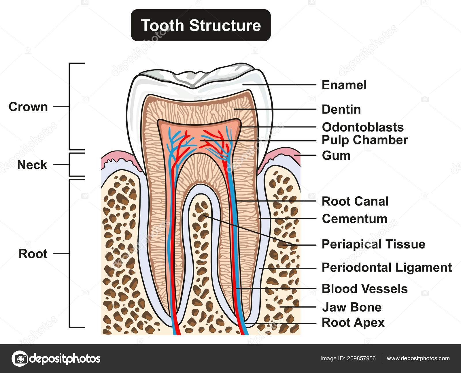
Labeled Tooth Cross Section Anatomy All Parts Including Crown Neck Vector Image By C Udaix Vector Stock 209857956 from st4.depositphotos.com □ on examining a cross section of any bone, it is composed of two kinds of bony tissue: This page discusses the calculation of cross section properties relevant to structural analysis, including centroid, moment of inertia, section modulus, and parallel axis theorem. We can see there are two layers of compact bone here. (left) a schematic diagram illustrating the assembly of collagen fibrils and fibers and bone mineral crystals. In the last decade, considerable technological improvements have been made to repair damaged bones and tissue, such as bone cross sections with implants for microscopic examinations. Diagram with articular cartilage, marrow, spongy bone, medullary cavity, endosteum, diaphysis, and periosteum. Cross section of the long bone. Diagram with articular cartilage, marrow, spongy bone, medullary cavity, endosteum, diaphysis, and.
Explaned distal and proximal epiphysis.
Cross section of bone diagram. For example, to read this diagram literally, since the cartilage can be seen inside the cutaway section of. Diagram with articular cartilage, marrow, spongy bone, medullary cavity, endosteum, diaphysis, and periosteum. can be used for personal and commercial purposes. The cross section of this circular cylinder is a circle. Cross sections are usually parallel to the base like above, but can be in any direction. The periosteum contains many strong collagen fibers that are used to firmly anchor. Diagram with articular cartilage, marrow, spongy bone, medullary cavity, endosteum, diaphysis, and periosteum. (b) in this micrograph of the osteon, you can clearly see the concentric lamellae and central canals. This bone is located directly beneath the skin on the anterior aspect of the leg (top of the image). Bone is hard and many of its functions depend on that characteristic hardness. Cheek bone (zygoma) upper jaw. Explaned distal and proximal epiphysis. (left) a schematic diagram illustrating the assembly of collagen fibrils and fibers and bone mineral crystals.
Bone cross section for radius digital science on behance cross section of a bone. As a part of the.


0 Comments